
Rectus Femoris Transfer

Why is this surgery recommended?

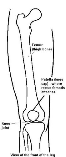
  • A tight or spastic rectus femoris (muscle on the front of your thigh) will cause the knee to be too straight (in extension) and a stiff knee during walking.
  • The surgery is done to improve knee bending (flexion) when it is limited by a tight or spastic rectus femoris muscle

What happens during the surgery?

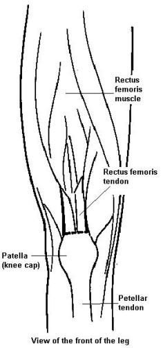
Your child's surgeon will make a cut at the front of the leg above the knee cap.

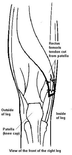
The rectus femoris tendon is detached from the top of the knee cap.

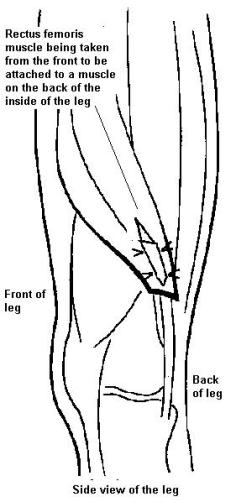
The tendon is then transferred and reattached to a muscle (semitendinosus) on the inside back of the leg, just above the knee.

This allows the rectus femoris muscle to bend (flex) the knee instead of straighten (extend) the knee.

Diagram of leg bones from the front, showing femur, patella and knee joint

Diagram of leg bones from the front, showing rectus femoris muscle, rectus femoris tendon, patella and patellar tendon

Diagram of front of the right leg, showing rectus femoris tendon cut from the patella

Diagram of leg bones from the side, showing rectus femoris muscle being taken from the front to be attached to a muscle on the back of the inside of the leg

(Illustration by Gillette Children's Specialty Healthcare)


SOURCE: Rectus Femoris Transfer ( )
Page printed: . Unofficial document if printed. Please refer to SOURCE for latest information.

Copyright © BC Children's Hospital. All Rights Reserved.

    Copyright © 2024 Provincial Health Services Authority.